3. Requesting Specific Imaging
MRI or Arthro-MRI are not systematic. Standard X-rays often provide sufficient information to establish an accurate diagnosis. In our experience, ultrasound is very rarely requested. We prefer printed imaging rather than CD-based imaging, which can sometimes be impossible to read depending on the software used.
I. The patient consults for pain that interferes with daily activities and prevents them from sleeping on their side
– Pain localised on the lateral side of the arm
This is likely a rotator cuff tendon injury. It may extend towards the neck and chest, suggesting a shoulder calcification. It may also be associated with pain on the anterior side of the arm, indicating inflammation or an associated injury of the long head of the biceps tendon.
– In all cases, we request an AP X-ray of the shoulder in neutral rotation and a lateral outlet view
- The AP X-ray reveals a shoulder calcification. The precise diagnosis of the cause of pain is established. Any additional imaging (MRI, ultrasound) is unnecessary.
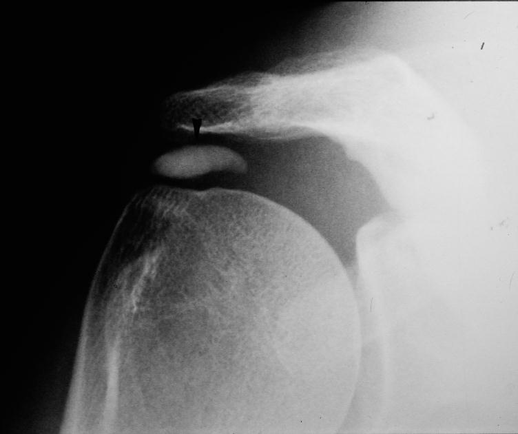
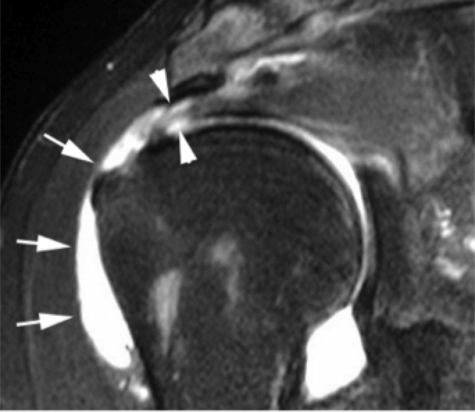
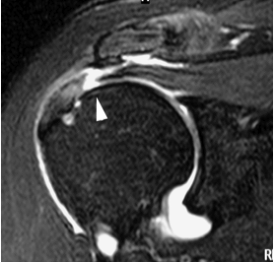
- The lateral outlet view reveals an aggressive anterior acromion that can cause damage to the rotator cuff tendons and the long head of the biceps tendon. In this case, Arthro-MRI (MRI with contrast injection) is necessary. This specific examination will reveal the condition of the cuff tendons: simple wear caused by the acromial bony spur, or a true tendon tear. Arthro-MRI is more precise than standard MRI. If the patient is claustrophobic, Arthro-CT scan can replace Arthro-MRI.