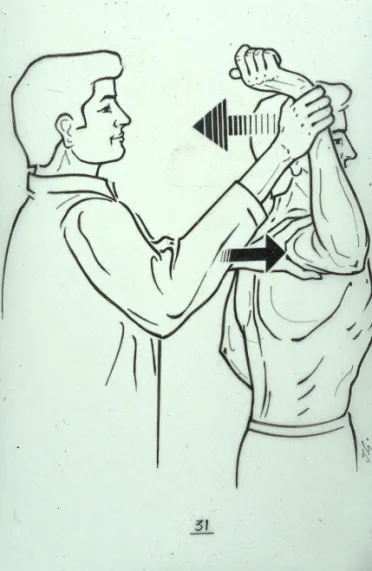
2. Examining the Patient
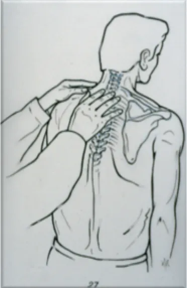
Both shoulders must be examined, undressed, along with the cervical and dorsal spine.
The patient being examined comes to consult either for pain, or because their shoulder dislocates.
I. The patient consults for pain
The first thing I say to the patient is: "Show me where your pain is."
Pain on the lateral side of the arm
This is probably a lesion of the rotator cuff tendons. I immediately look, through a comparative examination of both shoulders, for any limitation of mobility of the painful shoulder compared with the healthy one:
- Limited mobility in forward elevation
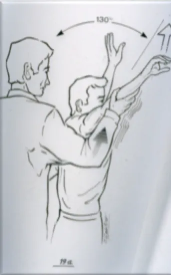
- Limited mobility in external rotation
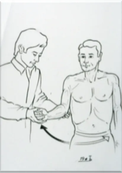
- Limited mobility in internal rotation, hand behind the back
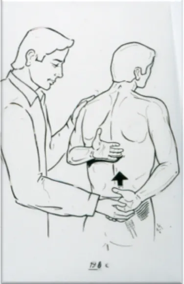
It is common for some degree of stiffness to be present, which amplifies the pain… A vicious pain-stiffness circle then forms, which is why early diagnosis matters.
Very specific clinical tests can determine, during the examination, whether one or more rotator cuff tendons are torn, caused by friction wear, age, and worsened by a possible fall onto the shoulder. Imaging will clarify the diagnosis.
Pain on the lateral side of the arm, radiating towards the neck and chest
If a cardiac problem has been ruled out, this is probably a shoulder calcification. Imaging will clarify the diagnosis.
Pain on the front of the arm
This is probably inflammation of the long head of the biceps, caused by repetitive movements (computer mouse, gardening, etc.).
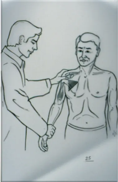
Pain on the acromioclavicular joint
This may be the after-effects of a minor fall or a sporting incident (combat sports, ice hockey, rugby, skiing, horse riding, cycling…), especially if there is an associated deformity. Imaging will clarify the diagnosis.

Pain localised in the shoulder with clicking whenever the patient moves their arm
- Arm mobility is reduced and painful. Most often, this is a patient over 65, already fitted with a hip or knee prosthesis.
- This is probably shoulder arthritis, known as omarthrosis.
- If the patient can raise the arm above horizontal, the rotator cuff tendons are presumably intact: this is called centred primary omarthrosis.
- If the patient cannot raise the arm, the rotator cuff tendons have probably disappeared, in a patient over 70: this is called eccentric omarthrosis with massive rotator cuff tear.
- In both cases, imaging will clarify the diagnosis.
Pain starting at the neck and radiating down the arm to the fingers
- This pain may be accompanied by tingling in the fingers. It is spontaneous or triggered by neck rotation or tilting movements: this is called cervico-brachial neuralgia, which can also be associated with rotator cuff lesions. This is why examination of the cervical spine must be systematic.
- Imaging will clarify the diagnosis.
II. The patient consults because their shoulder has dislocated several times
The first thing I look for on clinical examination is constitutional ligamentous hyperlaxity.
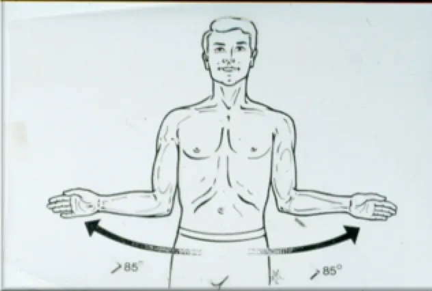
The patient's joints are overly flexible, and a history of repeated ankle sprains or knee pain should be sought. This ligamentous hyperlaxity translates, at the shoulder, into excessive external rotation exceeding 85 degrees. Ligamentous hyperlaxity worsens shoulder instability.


The clinical examination carefully looks for patient apprehension when the shoulder is placed in a dislocation-prone position, i.e. lateral elevation of the arm above horizontal combined with external rotation (the "cocked arm" position of a handball throw).
Imaging will clarify the diagnosis.