Requesting Specific Imaging
MRI or Arthro-MRI are not systematic. Indeed, "standard" X-rays often provide enough information to establish a precise diagnosis. In our experience, ultrasound is very rarely prescribed. We prefer imaging on paper rather than on CD, which is sometimes impossible to read depending on the software used.
I. The patient consults for pain that interferes with daily activities and prevents sleeping on that side
Pain localised on the lateral side of the arm
This is probably a rotator cuff tendon lesion. It may radiate towards the neck and chest: this could be a shoulder calcification. It may be associated with pain on the front of the arm: there is inflammation or an associated lesion of the long biceps tendon.
In every case, we request a neutral-rotation face X-ray of the shoulder and a cuff outlet profile X-ray.
- The face X-ray shows a shoulder calcification. The precise cause of the pain is identified. Further imaging (MRI, ultrasound) is unnecessary.
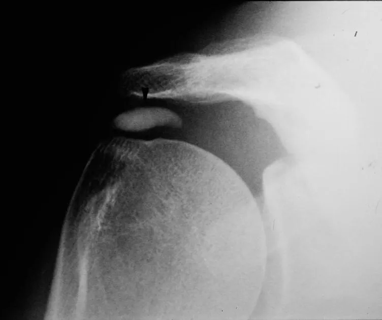
- The cuff outlet profile X-ray shows an aggressive anterior acromion that can cause rotator cuff and long biceps tendon lesions. This is when Arthro-MRI (MRI with contrast injection) is needed. Arthro-MRI is more precise than MRI. If the patient is claustrophobic, an Arthro-CT scan can replace the Arthro-MRI.
Pain on the acromioclavicular joint
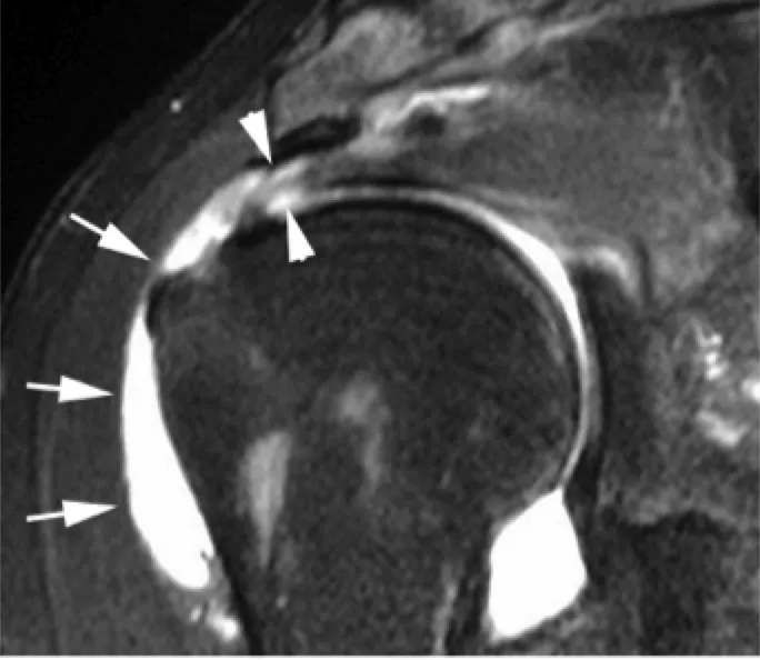
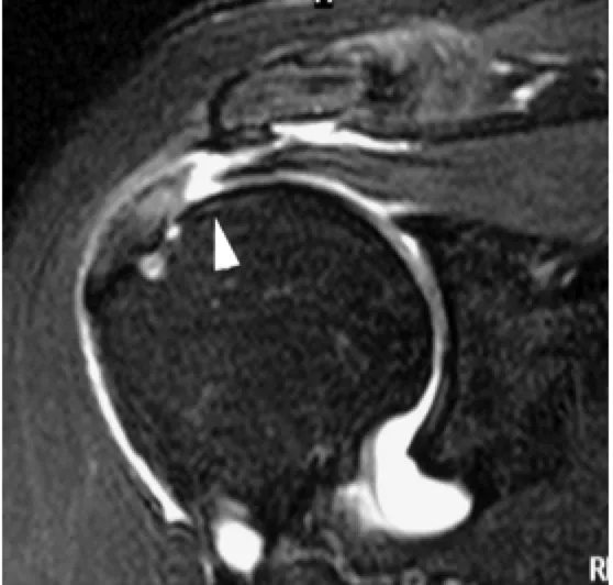
- Imaging is necessary to establish a precise diagnosis when there is no deformity after a fall. A comparative face X-ray of both shoulders can detect involvement of the outer end of the clavicle on the painful side.
Pain localised in the shoulder with clicking
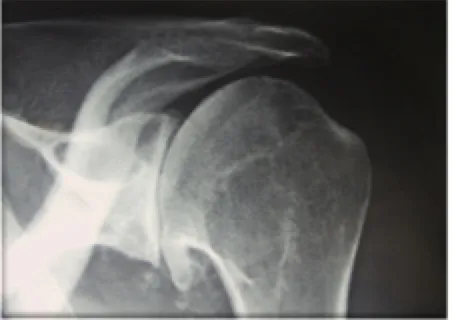
- Shoulder arthritis, or omarthrosis, is clinically suspected. In every case, we request a neutral-rotation face X-ray and an axillary profile view.
- The neutral-rotation face X-ray shows centred primary omarthrosis. The axillary profile provides information about the glenoid. An MRI will be needed to confirm the rotator cuff is intact, if a total shoulder prosthesis is considered.
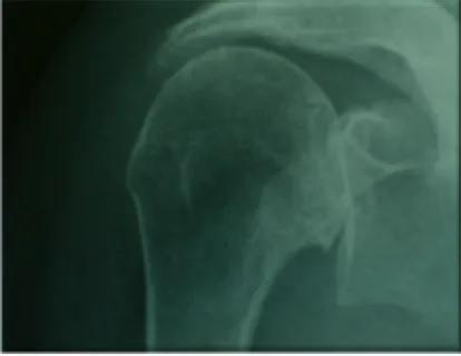
- The neutral-rotation face X-ray shows eccentric omarthrosis with a massive rotator cuff tear and the humeral head risen under the acromion. We know from experience that the rotator cuff tendons are torn and irreparable, especially if forward elevation is impossible with a complete loss of muscle strength. MRI is not necessary.
Pain starting at the neck and radiating down the arm with tingling in the hands
We refer the patient to a spine specialist, rheumatologist or surgeon, who will themselves request the specific imaging needed to establish the diagnosis: "standard" X-rays and cervical spine MRI.
II. The patient consults because their shoulder has dislocated several times
We request "standard" X-rays with two specific views that can, on their own, establish the diagnosis of anterior instability by showing characteristic bone lesions:
- The internal-rotation face X-ray can show a "notch" on the humeral head.
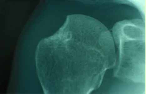
- The Bernageau profile view can show a "worn fracture" of the anterior glenoid rim. No further imaging is needed (CT scan, MRI).
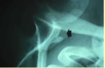
If "standard" X-rays show no bone lesion, particularly in a hyperlax patient, an arthro-CT scan is needed, which can establish the diagnosis of anterior instability by showing a Bankart ligament lesion.
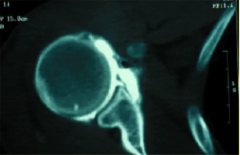
If the arthro-CT scan is normal, an arthroscopic examination of the shoulder under general anaesthesia will be needed to establish a precise diagnosis.